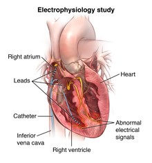
A cardiac electrophysiological study (EP study) is a minimally invasive procedure that is used to evaluate abnormal heart rhythm disturbances.
 During an EP study, small, thin wire electrodes are inserted through a vein in the groin (or neck in some cases). The wire electrodes are threaded into the heart, using a special type of X-ray, called fluoroscopy. Once in the heart, electrical signals are measured. Electrical signals are sent through the catheter to stimulate the heart tissue to try to initiate the abnormal heart rhythm.
During the EP study, doctors may also map the spread of electrical impulses during each beat. This may be done to locate the source of an arrhythmia or abnormal heart beat. If a location is found, an ablation (elimination of the area of heart tissue causing the abnormality) may be done.
The results of the study may also help the doctor determine further therapeutic measures, such as inserting a pacemaker or implantable defibrillator, adding or changing medications, performing additional ablation procedures, or providing other treatments.
Facts about Catheter Ablation
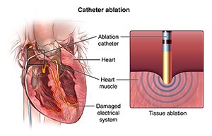
Also known as a cardiac ablation or radiofrequency ablation, this procedure guides a tube into your heart to destroy small areas of heart tissue that may be causing your abnormal heartbeat.
Not everyone with a heart arrhythmia needs a catheter ablation. It’s usually recommended for people with arrhythmias that can’t be controlled by medication.
The Procedure
 Before the procedure begins, you will be given intravenous medications to help you relax; some people even fall asleep. After the medication has taken effect, your doctor will numb an area on your groin and make a small hole in your skin. Then, the doctor will guide a thin guide wire and 2 to 3 small catheters through blood vessels to your heart. In some cases, your doctor may place several catheters, which are used to help guide the procedures.
After the catheter has been placed correctly, electrodes at the end of the catheter are used to stimulate your heart and locate the problem areas that are causing the abnormal heart rhythm. Various measurements of the electrical system are performed. If a person is in normal rhythm at the time of the procedure, an attempt is made then to reproduce the abnormal rhythm by pacing the heart through the catheters. Occasionally an intravenous medicine called isoproterenol is required to “rev up” the heart in order to reproduce the abnormal rhythm. Then, the doctor will use mild radiofrequency heat energy to destroy or “ablate” the problem area. This area is usually quite small, about one-fifth of an inch. Once the tissue is destroyed, the abnormal electrical signals that created the arrhythmia can no longer be sent to the rest of the heart. These radiofrequency lesions have no long-term adverse consequences. Overall cure rates with catheter ablation is >90% and can be as high as 96-99% depending on the specific type of rhythm problem.


